Figure 3.
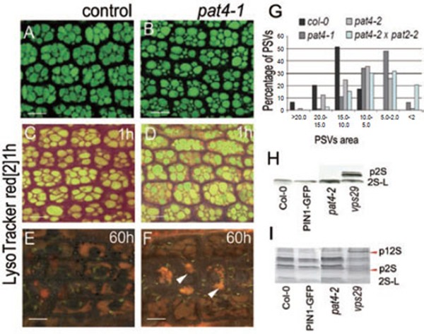
Defects in the transition between PSVs and lytic vacuoles, but not in trafficking to the PSVs in pat4 mutants. (A-B) PSVs in embryo root cells of control (A) and pat4-1 (B). (C-F) Accumulation of LysoTracker red following 1 h (C, D) and 60 h (E, F) of imbibition in wild-type (C, E) and pat4-1 (D, F) seeds, indicating defects in the acidification of mutant PSVs. (G) Quantification of the PSVs morphology. pat4-1 mutant (gray bar), pat4-2 mutant (light gray bar) and double mutant pat4 × pat2 (light blue bar) show smaller PSVs compared to wild-type seeds (black bar). Western blot (H) and SDS-PAGE (I) revealed no mis-secretion of 2S albumin and 12S globulin precursors in pat4-2 mutant but accumulation in the vps29, a mutant known to mis-sort reserve proteins. Scale bar = 10 μm.

