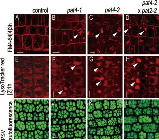
Figure 5.
Vacuole morphology in pat4-2 × pat2-2 double mutants. (A-D) FM4-64 uptake (3 h) in control (A), pat4-1 (B), pat4-2 (C) and pat4-2 × pat2-2 double mutant (D). Similarly to the single pat4 mutants, the vacuole morphology in the double mutant was impaired (arrowheads). (E-H) LysoTracker red accumulation in control (E), pat4-1 (F), pat4-2 (G) and pat4-2 × pat2-2 double mutant (H) showed abnormal acidification and lytic vacuole morphology in the single and double mutants (arrowheads). (I-L) PSVs autofluorescence imaging revealing defective PSVs morphology in single pat4-1 (J), pat4-2 (K) and pat4-2 × pat2-2 double mutants (L) as compared to wildtype embryos (I). Scale bar = 10 μm.

