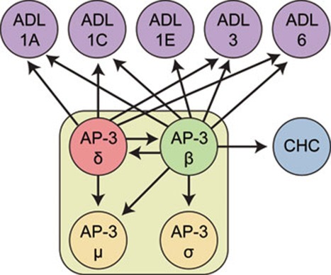
Figure 7.
Schematic representation of AP-3 δ and AP-3 β interacting proteins. Immunoprecipitations using both, AP-3 δ-GFP and AP-3 β-GFP as baits identified interaction with the remaining μ and σ subunits of the AP-3 complex, as well between AP-3 δ and/or AP-3 β and Arabidopsis dynamin-like (ADL) and clathrin heavy chain (CHC) proteins.

