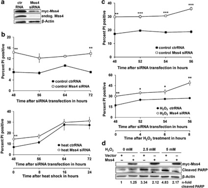
Figure 4.
Mss4 downregulation induces cell apoptosis. (a) A7 melanoma cells stably overexpressing human myc-tagged Mss4 were transfected with control or Mss4 specific siRNA for 48 h and the knockdown efficiency of endogenous and recombinant Mss4 protein was evaluated by Western blotting. Immunoblot for β-actin served as loading control. (b and c) A7 melanoma cells stably overexpressing human myc-tagged Mss4 were transfected with control or Mss4 specific siRNA. Forty-eight hours later the cells were either stimulated with 42 °C (b) or 5 mM H2O2 (c) for times indicated, stained with PI and the number of apoptotic cells was determined by flow cytometry. Mean values±S.D. from 2–3 repeated experiments are shown. (d) A7 melanoma cells were transiently transfected with myc-tagged Mss4 or empty vector for 40h. Then the cells were stressed with 2.5 or 5 mM H2O2 for 8h, lysed with RIPA buffer and the expression of Mss4 protein, as well as cleaved PARP was evaluated by western blotting. The β-actin immunoblot served as loading control. The n-fold PARP cleavage was estimated densitometrically as the relative intensity of cleaved PARP bands to the loading controls. Values of vector transfected and non-stressed cells were taken as unity. All experiments were carried out at least three times. *P<0.05, **P<0.01 and ***P<0.001 relative to ctrRNA transfected unstressed cells, t-test

