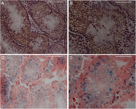
Figure 2.
Immunostaining with anti-GFP showed that T2R5 was expressed in early to mid-stage round spermatids, but not in spermatocytes or spermatogonia (A and B) in T2R5-CreGFP transgenic mice. (A) Low magnification. (B) High magnification from dotted frame in (A). After X-gal staining, positive signals were also observed in spermatids (C and D). (C) Low magnification. (D) High magnification from dotted frame in (C). Scale bar: 50 μm.

