Figure 1.
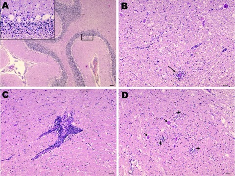
Nonsuppurative encephalitis in goat affected by louping ill. A) Cerebellum with necrosis of Purkinje cells. Hematoxylin and eosin (H&E) stain; scale bar = 100 µm. Inset: necrosis of Purkinje cells. H&E stain; scale bar = 20 µm. B) Midbrain. Area of neurophagia (arrow) surrounded by microglial cells. Necrosis of neurons can be also seen. H&E stain; scale bar = 50 µm. C) Lymphoid perivascular cuff in midbrain. H&E stain; scale bar = 50 µm. D) Spinal cord, gray matter. Focal microgliosis (crosses) and neurons undergoing necrosis (arrows). H&E stain; scale bar = 50 µm.

