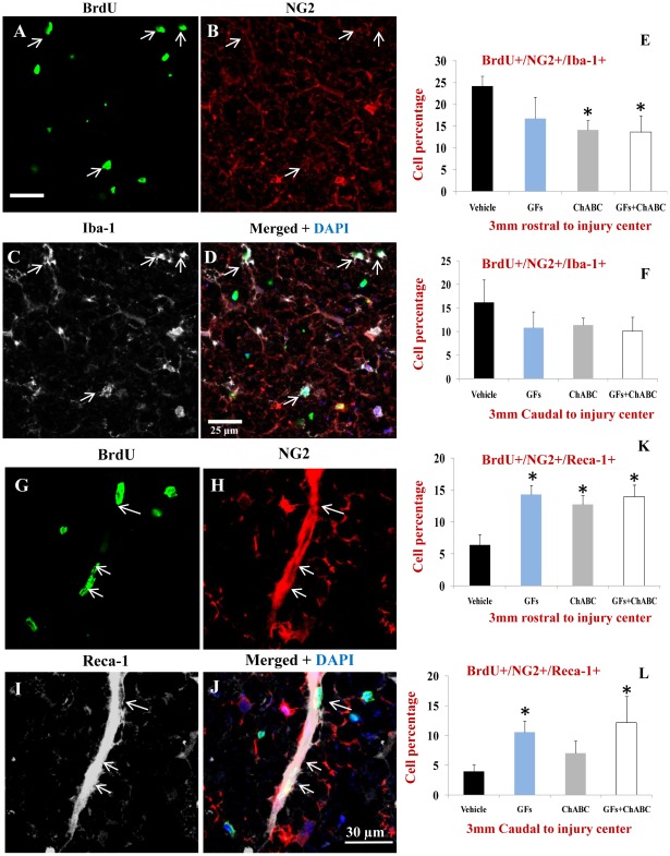
Figure 7. ChABC and GF treatments attenuate the proliferation of microglia/macrophages and promote the generation of new endothelial cells after SCI.
(A–D) Representative confocal images of BrdU+/NG2+ macrophages/microglia marked with Iba-1 in the injured spinal cord (arrows). (E–F) Under baseline SCI condition, macrophages/microglia comprised about 25% and 17% of BrdU+/NG2+ cells in rostral and caudal points to the injury center, respectively. After treatment with ChABC and/or GFs, we found a reduction in the number of BrdU+/NG2+/IbA-1+ cells that was statistically significant for ChABC and ChABC+GFs treatment groups relative to the vehicle group. (G–J) Representative confocal images show newly generated endothelial cells marked by Reca-1 and NG2 among BrdU+ cells. Reca-1 positive endothelial cells comprised a subpopulation of proliferating NG2+ cells after SCI (J). (K–L) Quantification of BrdU+/NG2+/Reca-1+ cells showed a significant number of newly generated endothelial cells after treatment with ChABC and/or GFs at both rostral and caudal points to the injury center compared to the vehicle group. *p<0.05, n = 6/group.

