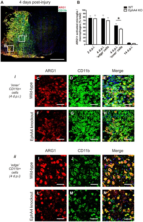
Figure 5. The proportion of Arginase 1-positive macrophages/activated microglia is lower in EphA4 knockout compared to wild-type spinal cords at 4 days post-injury.
Transverse sections of hemisectioned spinal cord were double-labelled with Arginase 1 and CD11b to identify the subset of infiltrating macrophages/activated microglia with Arginase 1 immunoreactivity. A representative example from a wild-type spinal cord at 4 days post injury is shown in A, with positive staining on the injured left hand side. Cells with a foamy macrophage appearance located in areas of tissue necrosis (ii, coded ‘edge’ areas) were more likely to be strongly Arginase 1-immunoreactive than densely packed macrophages not surrounded by obvious areas of necrosis (i, coded ‘inner’ areas). There was a significantly lower proportion (*p = 0.0018, B) of Arginase 1-positive CD11b positive macrophages/activated microglia in densely packed ‘inner’ areas in EphA4 knockout (F-H) compared to wild-type (C-E) spinal cord lesions at 4 days but no difference in the proportion (B) of Arginase 1-positive macrophages/activated microglia in necrotic ‘edge’ areas in EphA4 knockout (L-N) and wild-type (I-K) lesions. There was no difference between genotypes in the proportion of Arginase 1-positive macrophages/activated microglia at 2 or 14 days post-injury (B), by which time the proportion of Arginase 1-positive macrophages/activated microglia had dropped dramatically. Merged images include DAPI nuclear counterstain. Results show mean ± SEM of n = 6 per genotype at 4 days and n = 3 per genotype at 2 days and 14 days post-injury Scale bars: A = 500 µm; C-E, F-H, I-K, L-N = 50 µm. ARG1 = Arginase 1, WT = wild-type, KO = knockout, d.p.i. = days post-injury.

