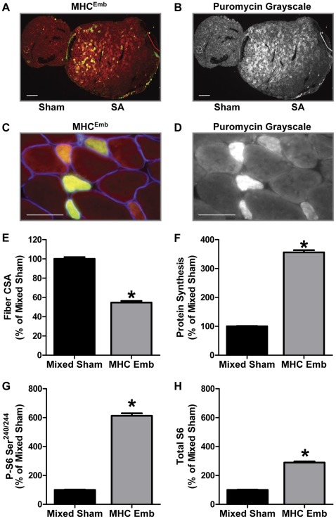
Figure 5. Cross-Sectional Area, Protein Synthesis, Ser240/244 Phosphorylated and Total Ribosomal S6 Protein in MHCEmb Positive Fibers.
Plantaris muscles obtained from control (Sham) and 10 d synergist ablated (SA) mice were frozen adjacent to one another, cross-sectioned, and then subjected to immunohistochemistry for MHCEmb and rates of protein synthesis (puromycin), Ser240/244 phosphorylated S6 (P-S6 Ser240/244), or total S6, as described in Figures 2 and 4. (A) Representative image of the signals for puromycin (red) and MHCEmb (green). (B) Grayscale image of the puromycin signal shown in A. (C) Higher magnification image from a SA muscle that was tripled stained for puromycin (red), MHCEmb (green) and laminin (blue). (D) Grayscale image of the puromycin signal shown in C. (E) The relative cross-sectional area (CSA), (F) rate of protein synthesis, (G) amount of P-S6 Ser240/244 and (H) total amount of S6 in MHCEmb positive fibers of SA muscles expressed relative to randomly selected fibers from sham muscles (Mixed Sham). The bars in A and B indicate a length of 200 μm, and the bars in C and D indicate 50 μm in length. All values are presented as the mean + SEM (n = 152–360 fibers / group from 6 independent pairs of muscles). ∗ Significantly different from mixed sham, (P<0.05).

