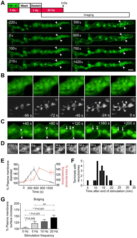
Figure 1. Bulging and collapsing of the presynaptic plasma membrane paves the way for bulk endocytosis.
NMJ preparations were pre-labeled with FM1-43 by electrical stimulation (1 Hz for 5 min), and extensively washed in normal Ringer's solution. Labeled nerve terminals were then stimulated at 20 Hz for 10 min and visualized by time-lapse confocal imaging starting from the final 4 min of stimulation. The data shown are from a representative experiment, repeated independently 14 times. (A) Representative time-lapse confocal images of nerve terminals at various time points during and after stimulation (time = 0 sec indicates the end of the 20 Hz stimulation period). Note the various remodeling stages: the progressive increase in plasma membrane diameter (bulging, an example of a prominent section is indicated by arrowheads), the rapid increase in intraterminal fluorescence following the collapse in plasma membrane diameter, and the appearance of ‘bulk endosomes’. (B and C) Magnified regions of the same nerve terminal in (A) showing the initiation of bulk endosomes as FM1-43-positive balls interconnected by growing tubules either during (B) or after (C) the stimulation period (arrows and arrowheads indicate formation of FM-labeled tubules). Bottom panel in (B) shows over-contrasted versions of images in the top panel to improve signal-to-noise ratios. (D) Time-course of the increase in internal diameter of a representative bulk endosome during the maturation process. (E) Graph showing the percentage increase in plasma membrane diameter (black, n = 5) and percentage increase in intraterminal fluorescence (red, n = 8 region of interests) plotted over time. (F) Histogram of time taken for formation of recyclosomes in nerve terminals stimulated at 20 Hz. (G) Stimulation frequency dependency of nerve terminal bulging. Data shown as mean ± S.E.M and statistic significance was determined using Student's t test. Scale bar 5 µm.

