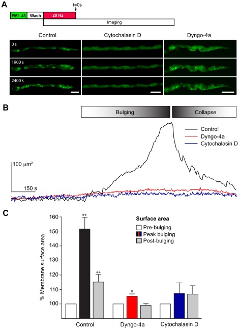
Figure 5. Disruption of actin polymerization and dynamin function inhibits plasma membrane bulging in nerve terminals stimulated at 20 Hz.
NMJ preparations were pre-treated with cytochalasin D (4 µM) or dyngo-4a (30 µM) for 40 min followed by a 5 min pulse with FM1-43 (5 µM). The preparation was washed in the continuing presence of cytochalasin D or dyngo-4a prior to electrical stimulation at 20 Hz for 10 min and time-lapse imaging. The data shown are from representative experiments, repeated independently 3–14 times. (A) Untreated nerve terminals displayed a bulging of the plasma membrane followed by collapsing and formation of recyclosomes. Treatment of nerve terminals with cytochalasin D or dyngo-4a blocked the plasma membrane bulging and formation of recyclosomes. (B) Typical dynamics of changes in the plasma membrane surface area in untreated (black), cytochalasin D-treated (blue) or dyngo-4a-treated (red) nerve terminals. (C) Statistical analysis of the changes in the estimated surface area of presynaptic plasma membrane at the peak of bulging and after the collapsing phase. Untreated nerve terminals showed a clear bulging and collapsing in response to 20 Hz stimulation, whereas dyngo-4a-treated terminals showed a limited, but significant bulging in comparison. No detectable change was detected in cytochalasin D-treated nerve terminals. Data shown as mean ± S.E.M and statistical significance was determined using Student's t test. Scale bars 5 µm.

