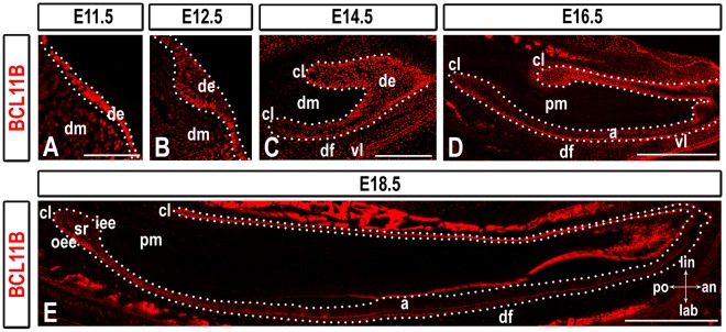
Figure 1. BCL11B expression during incisor development.
BCL11B immunostaining in sections of wild-type mice at indicated developmental stages. The epithelium is outlined by white dots. Scale bars: (A-B) 100 µm; (C) 200 µm; (D-E) 500 µm. a, ameloblasts; an, anterior; cl, cervical loop; de, dental epithelium; df, dental follicle; dm, dental mesenchyme; iee, inner enamel epithelium; lab, labial; lin, lingual; oee, outer enamel epithelium; pm, papillary mesenchyme; po, posterior; sr, stellate reticulum; vl, vestibular lamina.

