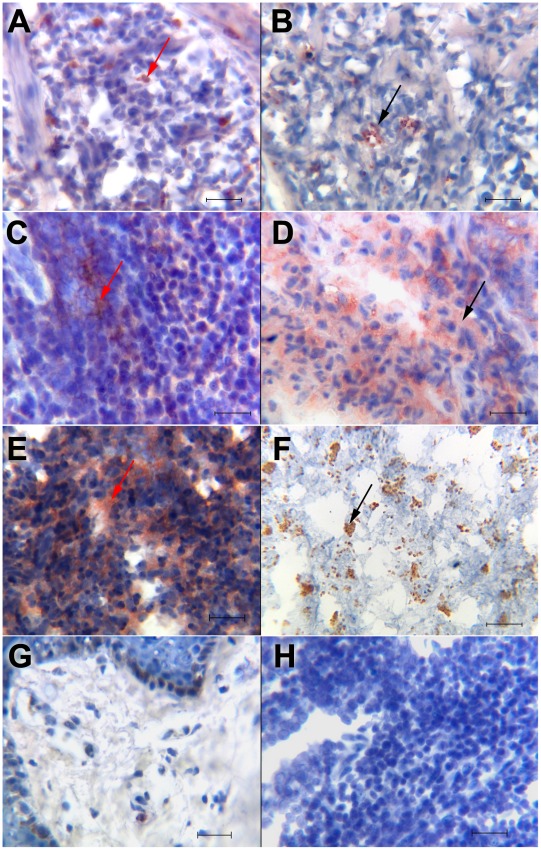
Figure 3. Immunohistochemistry for IL-1β and Leishmania mexicana staining in lesions of patients with cutaneous leishmaniasis.
(A) IL-1β staining on cells of LCL patient with single small lesion; (B) small clusters of Leishmania parasites in LCL patient with small ulcer; (C) diffuse distribution of IL-1β in LCL patient with abundant ulcers; (D) disintegrated Leishmania in LCL patient with abundant ulcers; (E) diffuse distribution of IL-1β in DCL patient; (F) clusters with abundant intact Leishmania parasites in DCL patient. Red arrows show IL-1β+ staining and black arrows show L. mexicana staining. (G) Normal skin was used as negative control for IL-1β immunostaining. (H) Control staining with secondary antibody. All sections were counterstained with haematoxylin. (A–H) scale bar = 50 µm. Immunostaining in tissue sections was visualized at a magnification of 400×. We show a representative result of different types of lesions within each group: LCL patients with one small ulcer: (n = 8 for IL-1β staining and n = 17 for L. mexicana staining) (A and B); LCL patients with various ulcers (n = 3) (C and D); DCL patients (n = 6) (E and F).

