
Figure 5.
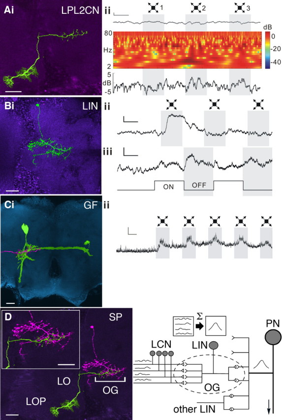
Neural integration enhances sensitivity to looming stimuli. A–C, Left, Confocal images of recorded neurons. Scale bars, 20 μm. Right, Corresponding recordings. Calibration: 2 mV/500 ms. Aii, Power spectrum analysis illustrates the nonspiking LPL2CN responding to the looming stimuli 2 and 3. The first trace is the recording sample. The time–frequency plot in the middle shows the power of membrane potential oscillations calculated from the recording sample above. The line plot at the bottom shows averaged powers (2–80 Hz) throughout the stimulus calculated from the time–frequency plot above. B, The unambiguous and rapidly adapting responses to looming and full-field flicker stimuli of the LIN in the GF glomerulus. C, The GF and its depolarizing response to looming stimuli. An image of the terminal of LPL2CN (pink) is superimposed on the GF dendrites to indicate their overlap in the GF glomerulus. D, Convergent processing in the optic glomerulus. Left, Montage showing overlap at the same optic glomerulus [bracketed optic glomerulus (OG)] of the recorded LIN (pink) and the axon terminal of a recorded LPL (green). This species of neuron, LPL2CN, belongs to the class of LPLs characterized by their dendrites in the lobula (LO) and lobula plate (LOP). Inset, Enlargement of the related glomerulus. Right, Schematic to illustrate convergence of LCNs to an OG. Responses of the LCNs are summed (Σ) and carried by the LIN relaying to its cognate projection neuron (PN). PNs of OG receive additional LIN inputs.