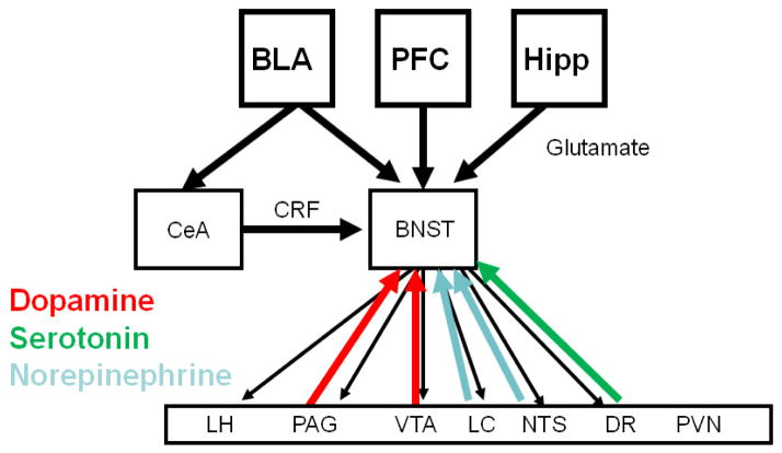
Figure 1.
Network diagram outlining the connectivity of the BNST. The BNST receives glutamatergic projections from the basolateral amygdala (BLA), the prefrontal cortex (PFC) and the hippocampus (Hipp) and a GABAergic/CRF projection from the central nucleus of the amygdala (CeA). The BNST then projects back to the CeA, as well as brain regions that mediate for the specific systemic and behavioral indicators of fear and anxiety. Shown on the diagram are the lateral hypothalamus (LH), periacqueductal grey (PAG), locus coeruleus (LC), nucleus tractus solitaries (NTS), ventral tegmental area (VTA), the paraventricular nucleus of the hypothalamus (PVN) and the dorsal raphe (DR). Several of these brain regions project back to the BNST, suggesting the possibility of feed-back and feed-forward circuits. It should be noted that the CeA also projects to the same targets as the BNST, however it does not project to the PVN.

