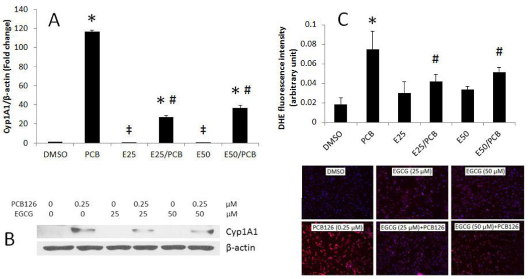
Figure 1.
EGCG attenuates PCB 126-mediated induction of Cyp1A1 and cellular oxidative stress. (A) Expression of mRNA was analyzed in endothelial cells pretreated with 25–50 µM of EGCG for 3 h, followed by treatment with PCB 126 at 0.25 µM for 4 h. Real-time PCR technique was used to measure expressed mRNA levels. (B) Expression of CYP1A1 protein in endothelial cells pretreated with 0–50 µM of EGCG for 3 h, followed by treatment with PCB 126 for 4 h using Western blot technique. Densitometry results were normalized to β-actin. The Western blot picture shown is a representative of three independent blots. Real-time PCR and Western blot results represent the mean ± SEM, with n=3. (C) Superoxide production in endothelial cells using DHE staining. DHE red fluorescence was assessed using fluorescence microscopy and the strength of signal was quantified. Experiments were repeated a minimum of three times. *Significantly increased compared to DMSO control. ‡Significantly decreased compared to DMSO control. # Significantly different compared to the PCB treatment group.

