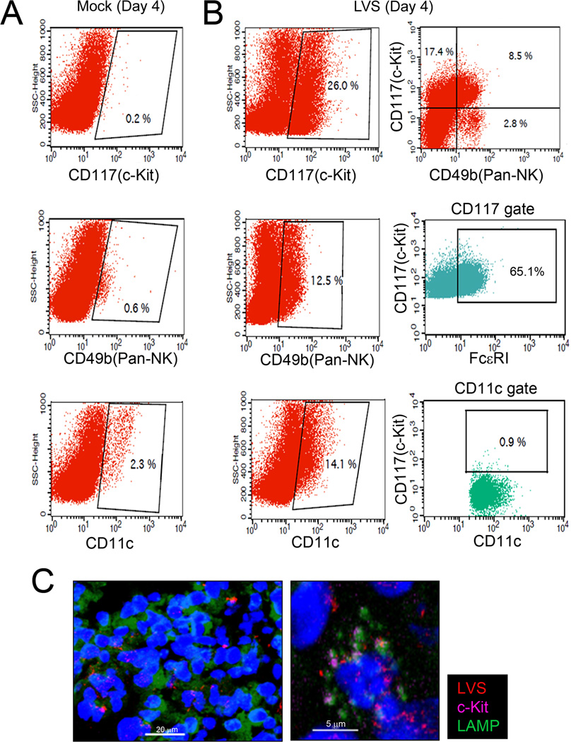
Figure 6. Mast cells infiltrate the lungs during pulmonary F. tularensis infection.
Lungs cells from WT and TLR2−/− mice were analyzed at days 2 and 4 post-challenge (day 4 shown). Cells were surfaced stained with cKit, CD49b (panNK cell marker), and CD11c (dendritic cell marker) fluorochrome conjugated antibodies or isotype control. (A) Day 4 Mock: representative scatter plots shown with c-Kit gate, NK cell gate, and dendritic cell gate. (B) Day 4 LVS infected lung cells: c-Kit gate, NK cell gate; and dendritic cell gates ; c-Kit+CD49b+ NK cells (top right dot plot) with further analysis within c-Kit gate (middle dot plot) for double positive c-Kit+FcεRI+ mast cells and c-Kit+CD11c+ dendritic cells (bottom right dot plot). Samples were acquired with FACS Calibur (50K–100K cells) and analyzed with CellQuest. Representative scatter and dot plots shown for mock and LVS infected lung cells. (C) Confocal microscopy analysis: c-Kit+LAMP+ cells (purple/green) in direct contact with mCherry LVS (KKF314); nucleus (DAPI, blue); representative confocal microscopy image (left) at 400X magnification with enlargement (right) shown.

