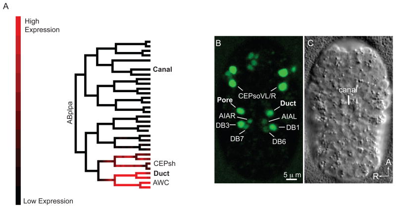
Figure 3. mls-2 is expressed in the duct and pore cell lineages.
(A) Lineage tree showing florescence intensity of GFP::MLS-2 expression from 3D automated lineage analysis. Only 1 of 3 embryos that were lineaged is shown here. Only the ABplpa lineage is shown, but GFP::MLS-2 is symmetrically expressed in the ABprpa lineage, which gives rise to the excretory pore. See Supplemental Fig.1 for complete lineage analysis of all 3 embryos. (B) Ventral enclosure embryo expressing GFP::MLS-2, and (C) corresponding DIC image. Identities of some nuclei are indicated. CEPsh nuclei are dim and not visible at this stage. AWC nuclei are not in plane of focus. The pair of nuclei directly above the CEPsoVL/R nuclei are the sisters of the CEPsoVL/R that are fated to die (Sulston et al., 1983). The DB1/DB3 ventral cord motor neurons are sisters of the duct and pore, respectively. AIAL/R are amphid inter-neurons and DB6/DB7 are ventral cord motor neurons; expression in these cells initiates at this stage and is very faint.

