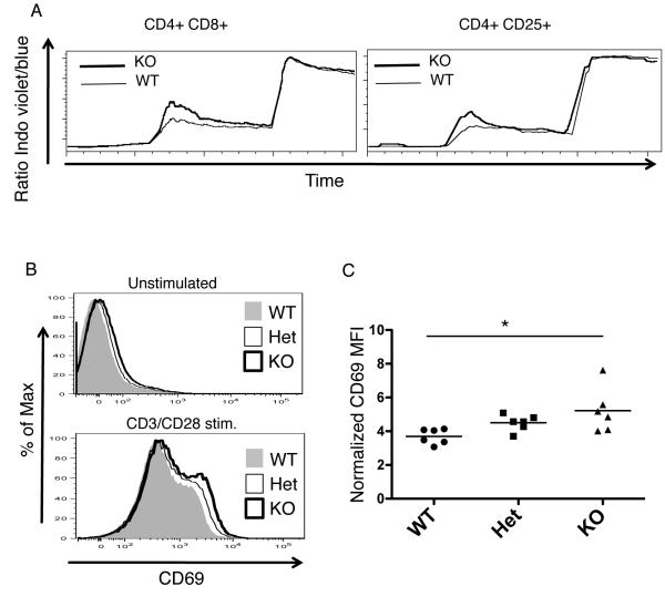
Figure 3.
Signaling differences in the thymus of PTPN22 deficient vs. WT mice. A, Calcium flux in double positive (DP) and CD4+CD25+ thymocytes derived from wild type (thin line) or KO (thick line) mice. Cells were pre-labeled with anti-CD3 biotin antibody and stimulated at 30 seconds with strepavidin. Exogenous calcium was added at 120 seconds and ionomycin at 300 seconds. For the experiment shown, WT cells were labeled with Cy5 and mixed with unlabeled KO cells to stimulate under identical conditions. During the same experiment the Cy5 labeling was reversed to ensure it did not interfere with Ca Flux. This is an example of one of 4 separate experiments (WT n=8 KO n=8). B. CD69 expression on DP thymocytes from WT (filled plot), Het (unfilled, thin line) and KO (unfilled, thick line) mice either from unstimulated thymocytes or CD3/CD28 stimulated thymocytes (overnight stimulation). C, Normalized MFI of CD69 on CD3/CD28 stimulated DP thymocytes. *, p< 0.05

