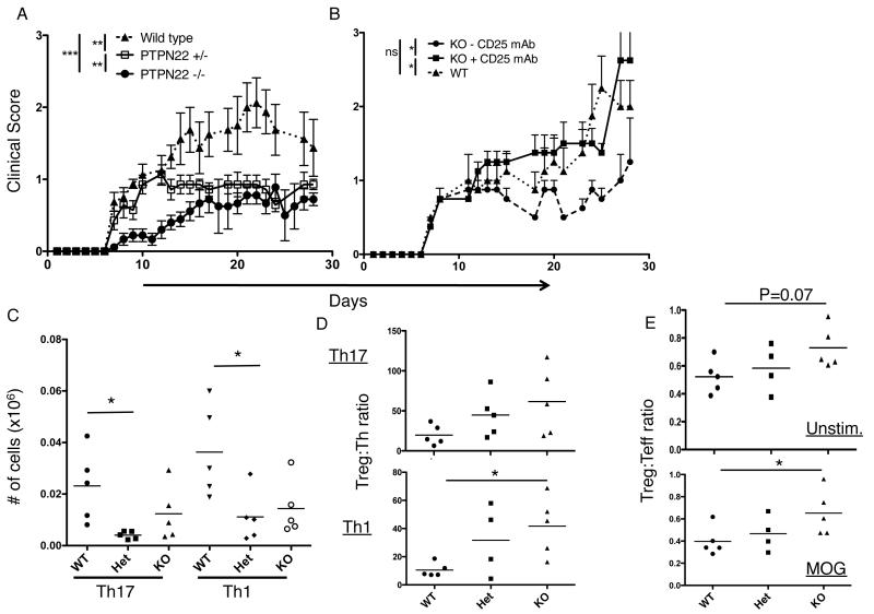
Figure 7.
PTPN22 deficient mice are protected from EAE. A. EAE was induced in female mice at 9 weeks of age by s.c. injection of MOG35-55 peptide in CFA followed by i.p. injection of pertussis toxin on days 1 and 2. Mice were scored each day for 28 days (n= WT 8, Het 7, KO 9). This experiment was performed twice with WT and KO mice n=4 . B. EAE was induced in WT and KO mice on day 1 as described and mice received an i.p. injection of anti-CD25 depleting antibody on days −1 and +4 (n= WT, 5; KO, 4; KO + CD25mAb, 4). C. EAE was induced as described in previously and draining lymph nodes were harvested at day 10 then restimulated with MOG for 5 hours then stained for IL-17 and IFN-γ. D. Harvested LN cells were restimulated with MOG peptide and stained for IL-17, IFN-γ and FoxP3, the Treg:Th ratio is plotted for both Th17 and Th1 cells. E. Harvested LN cells were stained immediately ex vivo (unstimulated, top panel) or restimulated with MOG peptide (bottom panel) as in part D. Treg/Teff ratio is plotted (effector CD4 cells are defined as CD4+ CD44hi Foxp3−). ***, p< 0.001; **p<0.01; *, p< 0.05

