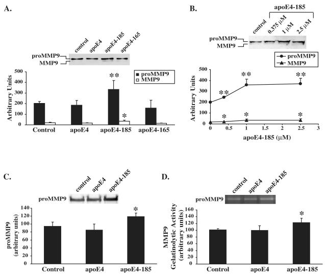
Figure 1A-C. Effect of WT and carboxy-terminal truncated apoE4 forms on MMP9 levels in SK-N-SH cells.
A, SK-N-SH cells were incubated in the absence (control) or presence of 1 μM lipid-free WT apoE4, apoE4-185 or apoE4-165 for 24 h. Secreted MMP9 levels were measured by immunoblotting as described under “Experimental procedures”. Western blots were scanned and quantified by ImageJ (lower panel). Values represent the means ± SD of six experiments performed in duplicate or triplicate. *, p = 0.01 vs. control. **, p < 0.005 vs. control. B, SK-N-SH cells were incubated in the presence of increasing concentrations of apoE4-185, as indicated, for 24 h. Secreted MMP9 levels were measured by immunoblotting. Western blots were scanned and quantified by ImageJ (lower panel). Values represent the means ± SD of four measurements. *, p < 0.05 vs. control. **, p < 0.005 vs. control. C, D, SK-N-SH cells transiently transfected with proMMP9 were incubated in the absence (control) or presence of 1 μM lipid-free WT apoE4 or apoE4-185 for 24 h. Secreted MMP9 levels were measured by immunoblotting (C) and enzymatic activity was assessed by gelatin zymography (D) as described under “Experimental procedures”. Western blots and zymograms were scanned and quantified by ImageJ (lower panel). Values represent the means ± SD of three experiments performed in duplicate. *, p < 0.05 vs. control.

