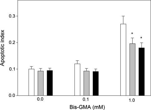
Fig. 7.
Apoptosis of human gingival fibroblasts exposed for 6 h at 37°C to the mixture of methacrylates containing 45% 2-hydroxyethyl methacrylate and 55% bisphenol A-diglycidyl dimethacrylate (w/w) (HEMA/Bis-GMA) at different Bis-GMA concentration without (white bars) or with a 1-hr incubation with 50 μM ascorbate (grey bars) or melatonin at the same concentration (black bars). Apoptosis was assessed by flow cytometry with Annexin V-FITC/propidium iodine. Apoptosis was expressed as a ratio of the number of early and late apoptotic cells to the number of cells with no evidence of apoptosis; *p < 0.05 as compared with unexposed control

