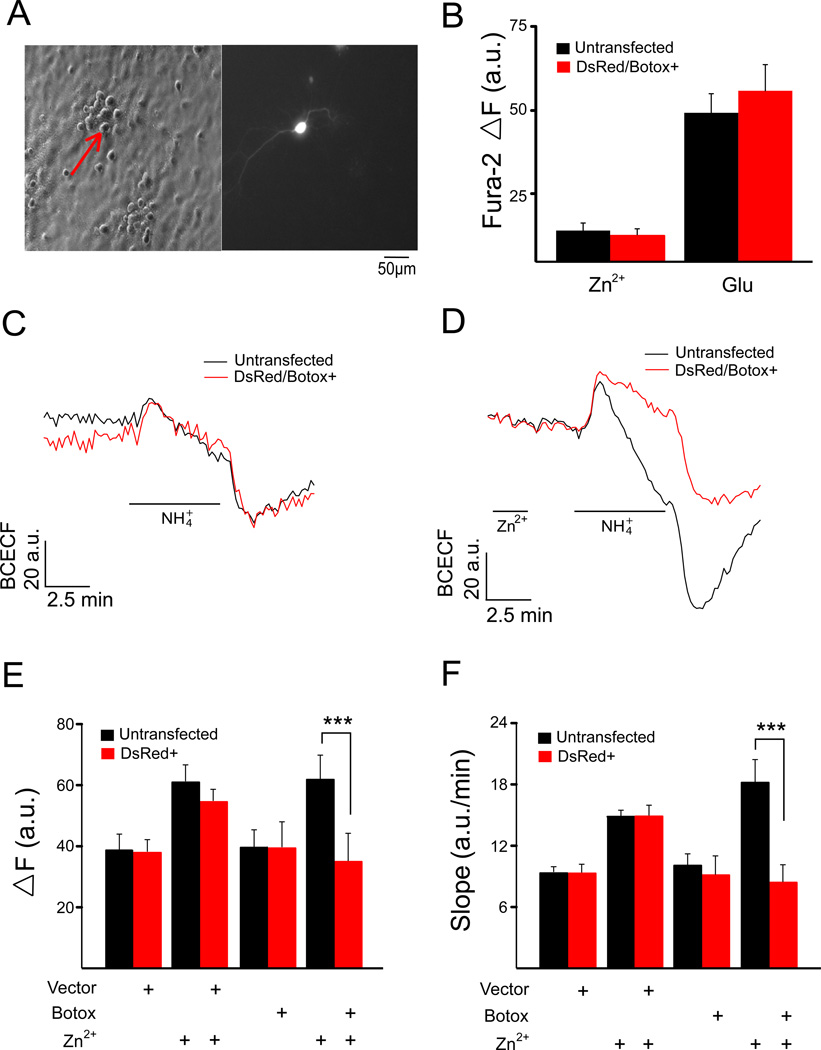
Figure 4.
KCC2 activity in botulinum toxin C1 (Botox C1)-expressing neurons. (A) Co-transfection with a DsRed expressing plasmid was used to locate Botox C1-transfected rat cortical neurons on coverslips. (B) Mean ΔF Fura-2 values (± SEM) for Ca2+ responses in untransfected and DsRed/Botox C1-expressing neurons from the same coverslips (n=3) treated with 200 µM ZnCl2 (Zn2+) and 300 µM glutamate (Glu). Fluorescence change of transfected (DsRed+ or DSRed/Botox C1+) and untransfected cells from the same coverslip in the absence (C) and presence (D) of a 2 min Zn2+ pretreatment. KCC2 activity, measured in terms of both ΔF (E) and slope (F) (mean ± SEM) for empty-vector and Botox C1 transfected rat cortical neuron, along with matched untransfected cells for each condition, in the absence and presence of Zn2+ pretreatment (n=6–9 coverslips per group). Zn2+-treated Botox C1-expressing cells showed significantly lower KCC2 activity when compared to the untransfected cells from the same coverslip; ***P<0.001 (paired t test).

