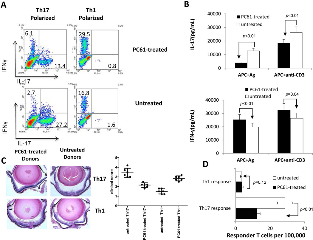
Fig. 1. Injection of B6 mice with anti-mouse CD25 antibody (PC61) decreases the Th17 autoreactive T cell response.
A). Splenic T cells from IRBP1–20/CFA-immunized mice with or without prior PC61 treatment were enriched by passage through nylon wool and stimulated for 48 h with an optimal dose of IRBP1–20 (10 µg/ml) under Th1 or Th17 polarized conditions and the activated T cells separated by Ficoll gradient centrifugation on day 3, then cultured under the same polarized conditions for a further 5 days and intracellularly stained with PE-conjugated anti-IFN-γ antibodies and FITC-conjugated anti-IL-17 antibodies, followed by FACS analysis.
B) IFN-γ and IL-17 levels in the culture supernatants after the 48 h incubation with peptide measured by ELISA.
C) IRBP-specific T cells (2 × 10⁶/mouse) from untreated and PC61-treated IRBP-immunized mice activated under Th1 or Th17 polarized conditions were adoptively transferred to syngeneic B6 mice. Pathologic examination was conducted 10 days after disease induction. A set of representative pathologic slides is shown along with summarized in vivo results.
D) Responder T cell numbers evaluated by LDA as detailed in the Materials and Methods.
The results shown are representative of those from >5 experiments.

