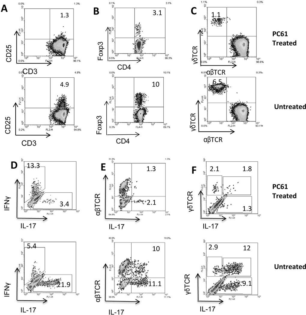
Fig. 2. Phenotypic change of T cells in PC61-injected mice.
B6 mice were randomly separated into two groups (n=8) and immunized with IRBP1–20/CFA with or without a prior treatment with PC61. After 13 days, splenic T cells were enriched and stimulated with the immunizing peptide. The phenotype of the T cells was analyzed either before (A–C) or after (D–F) in vitro stimulation.
A–C) Effect of PC61 injection on CD25+CD3+ cells (A), Foxp3+ cells (B), and γδ T cells (E).
D–F) After in vitro stimulation with the immunizing antigen and expansion under Th17 polarized conditions, the activated T cells were stained with the indicated antibodies and analyzed by flow cytometer. The experiments were repeated more than 5 times and the results of one representative experiment are shown.

