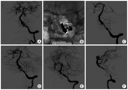
Fig. 4.
Follow-up angiograms at 4 months after final embolization also showing left VADA regrowth (A). Postoperative skull radiograph after telescopic stenting with two covered stents graft (Graftmaster 3.5×12 mm, 3.5×9 mm) on the left vertebral artery just below the PICA origin and a third stent (Driver 3.5×12 mm) across the PICA origin (B). Arrows indicate the proximal end of covered stent and distal end of Driver stent. Final vertebral angiograms at early arterial (C) and late arterial phase (D) still demonstrate slow retrograde flow to VADA, although flow velocity into the VADA is markedly decreased. Follow-up angiograms 2 month later show near complete occlusion of the left VADA (E and F). VADA : vertebral artery dissecting aneurysm, PICA : posterior inferior cerebellar artery.

