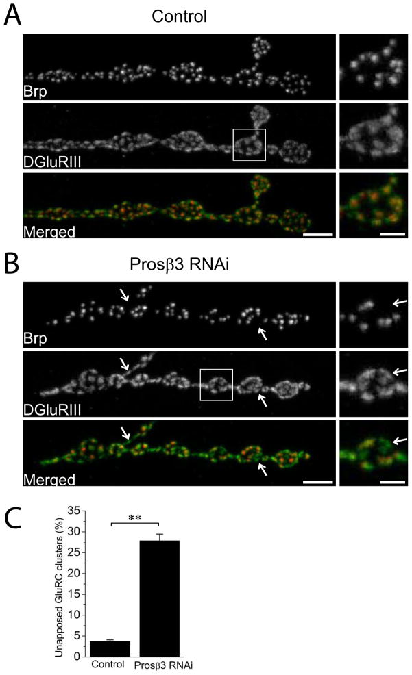
Figure 1. Neuron-driven RNAi targeting of a proteasomal subunit, Prosβ3, disrupts synaptic apposition.
Sample confocal images of muscle 4 NMJs of (A) Control (UAS-DCR2/+; Elav-Gal4/+) and (B) Prosβ3 RNAi knockdown (UAS-DCR2/+; UAS- Prosβ3 RNAi/+; Elav-Gal4/+ ) third instar larvae stained for the presynaptic active zone protein Brp (red) and the postsynaptic glutamate receptor DGluRIII (green). Inset shows a magnified image of boxed region to highlight the apposition of active zones and glutamate receptor clusters. Muscle 4 Type Ib NMJs from the genotypes in (A) and (B) were quantified for the percentage of unopposed DGluRIII clusters in C. Note that neuronal knockdown of Prosβ3 leads to significant increase in unapposed glutamate receptors (arrows); ** p<0.001; n = 16 for both genotypes. Error bars represent SEM. Scale bars represent 5 μm in lower magnification images and 2 μm in higher magnification inset.

