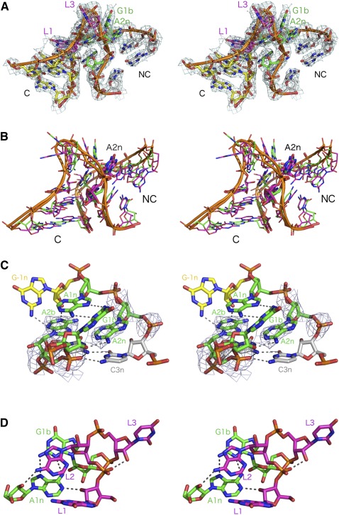
FIGURE 4.
The crystal structure of the SAM-I riboswitch in which the k-turn has been replaced by that of T. solenopsae Kt-23. Parallel-eye stereo views are shown. (A) The structure of the k-turn within the modified riboswitch. The 2Fobs-Fc electron density map is shown contoured at 1σ. The nucleotides are colored to match the schemes shown in Figure 1. (B) Superposition of Kt-23 (magenta) with the standard k-turn Kt-7 (green). The two k-turns were superimposed using their phosphorus atoms only. Note the close superimposition of A1n and A2b nucleobases (highlighted in stick form). (C) View from the nonbulged strand side of the core of the k-turn. The electron density shown on the A2b and A2n nucleotides is taken from the composite omit map. (D) View down onto the loop to show the conserved hydrogen bonds.

