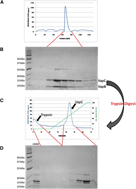
FIGURE 2.
Purification and isolation of VapC. (A) The VapBC complex Rv0065a-Rv0065 from M. tuberculosis expressed in M. smegmatis elutes as a single peak on an S200 size exclusion column. (B) The corresponding SDS-PAGE gel shows the presence of both VapB and VapC proteins in the complex. (C) Trypsin digestion of the VapBC complex followed by anion exchange chromatography. Due to differences in their isoelectric points, trypsin is eluted early from the column while VapC is eluted later in the NaCl gradient. (D) Corresponding SDS-PAGE gel demonstrates the trypsin digest of VapBC loaded onto the column (LOAD) and the presence of VapC with and without the C-terminal His-tag.

