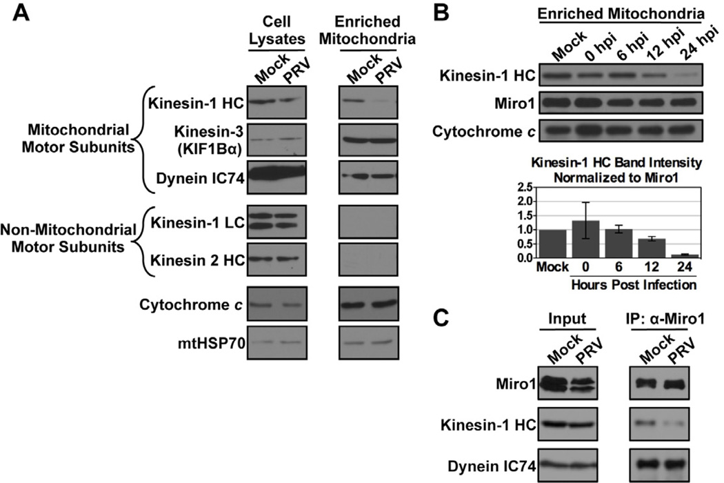
Figure 5. Western blot analysis of mitochondrial motor protein subunits during PRV infection.
(A) Mitochondria were enriched from PC12 cells that were mock or PRV Becker infected at 18 hpi. Kinesin-1 heavy chain (HC), dynein intermediate chain IC74, and the kinesin-3 KIF1Bα are known mitochondrial motor protein subunits. Kinesin light chain (KLC) and kinesin-2 are non-mitochondrial motor protein subunits and serve as controls for the purity of the mitochondrial enrichment preparation. Cytochrome c and mitochondrial HSP70 (mtHSP70) are loading controls. (B) Mitochondria were enriched from mock or PRV infected PC12 cells at the indicated times post infection. At each time point, the levels of kinesin-1 HC were normalized to those of Miro1, which recruits kinesin-1 HC to the mitochondrial surface. The mean of two biological replicates is shown with bars representing the range. (C) Lysates of mock or PRV infected PC12 cells at 18 hpi were subjected to co-immunoprecipitation (IP) using anti-Miro1 antibodies. The input (left panels) and the precipitate (right panels) were probed using antibodies against Miro1, kinesin-1 HC, and dynein IC74.

