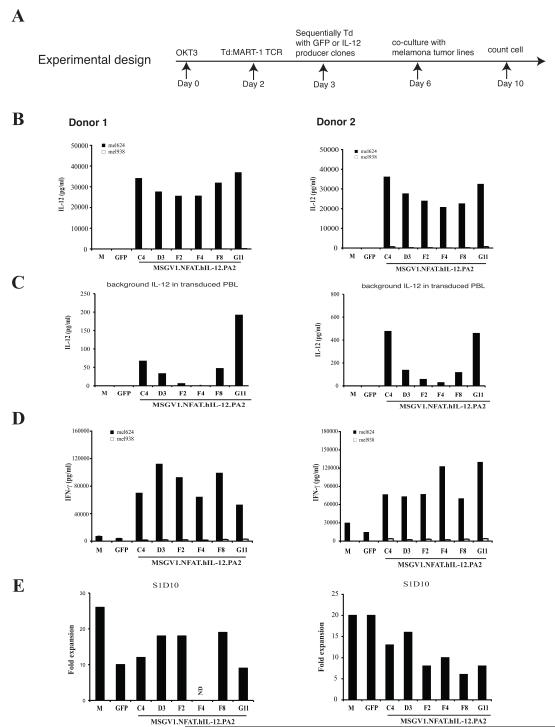
Figure 4. Screening NFAT.hIL12 viral producing clones in human PBL.
(A). The experimental design. PBLs were stimulated with soluble OKT3 at day 0. On day 2, the cells were suspended in fresh medium with IL-2 and transduced with MART-1 TCR. Next day, the MART-1 transduced cells were divided into 7-groups, and transduced with vector supernatant from six MSGV1.NFAT.hIL-12.PA2 vector producer clones: C4, D3, F2, F4, F8, G11 or GFP respectively. Three days after the 2nd transduction (day 6), the transduced cells were co-cultured with HLA-A2+/MART-1+ tumor line: mel624 and HLA-A2-/MART-1+ tumor line: mel938. The levels of IL-12 (B & C) and IFN-γ (D) in the culture were measured by ELISA (shown are the mean values of duplicate determinations). (E). The number of viable cells in different cultures was enumerated on day 10 by trypan blue staining. The data was repeated using PBLs from two donors. M, MART-1 TCR transduced.

