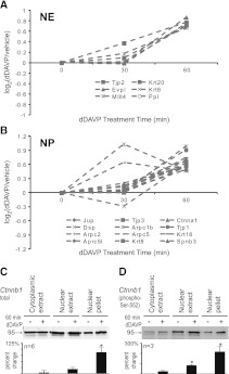
Figure 7.
Cytoskeletal and junctional proteins increased in nucleus. Values for cytoskeletal and junctional proteins in (A) nuclear extract fraction and (B) nuclear pellet fraction quantified at 30 minutes after dDAVP addition (0.1 nM) compared with values at 60 minutes of dDAVP addition. Eighteen of 20 of these proteins showed positive values at 30 min (P<0.01), chi-squared analysis versus random distribution of positive and negative (10/20). (C) Immunoblotting for total β-catenin in samples from three cellular fractions after 60 minutes of dDAVP (0.1 nM) or vehicle. (D) Immunoblotting for β-catenin phosphorylated at Ser-552. Results of densitometric quantification are shown in the bar graphs below the representative bands. *P<0.05 (t test). NE, nuclear extract; NP, nuclear pellet.

