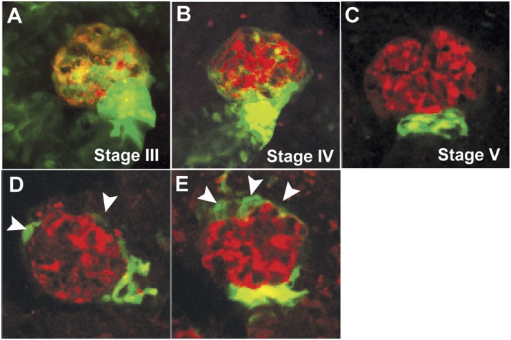
Figure 6.
Postinjury response in zebrafish mesonephric glomeruli. (A–C) The distribution of wt1b::GFP-expressing cells (green) during the different developmental stages of glomeruli in adult zebrafish23 resembles that of renal progenitors in adult human glomeruli.26 With the maturation of the nephron, wt1b::GFP expression is diminished in the epithelial cells except at the urinary pole of the Bowman capsule. The podocytes (red) are labeled with mCherry fluorescence. (D and E) After MTZ-induced podocyte injury, wt1b::GFP-expressing cells (arrowheads) are expanded toward the vascular pole of the Bowman capsule of mesonephric glomeruli in zebrafish, suggesting a podocyte repair mechanism similar to that in mammals.

