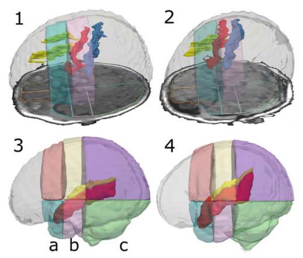
Fig. 3.
3D Models of Segmented Gyri and Sulci in Relation to Parcel Borders. The upper row shows 3D models of the superior frontal sulcus (yellow), the precentral sulcus (red), and the central sulcus (blue) inside a model of the ICC. The sulci segmentations are overlaid by transparent models of the superior precentral parcel (bright blue) and the superior central parcel (rose). An axial SPGR slide with an outline of the parcellation is also to be seen. Image 1 displays a preterm infant’ segmentation, image 2 displays a fullterm case. Two representative cases were chosen to visualize how the precentral sulcus is almost entirely shifted into the central region in preterm infants. The lower row shows 3D models of the superior temporal gyrus (red) and the Sylvian fissure (yellow) overlaid by 3D models of the superior and inferior precentral (a), central (b) and occipital parcels (c). Image 3 displays a preterm case, image 4 a fullterm case. Note how in the preterm infant, the superior temporal gyrus and the Sylvian fissure are shifted downwards and show a flat orientation with regard to the parcellation in comparison to the fullterm infant.

