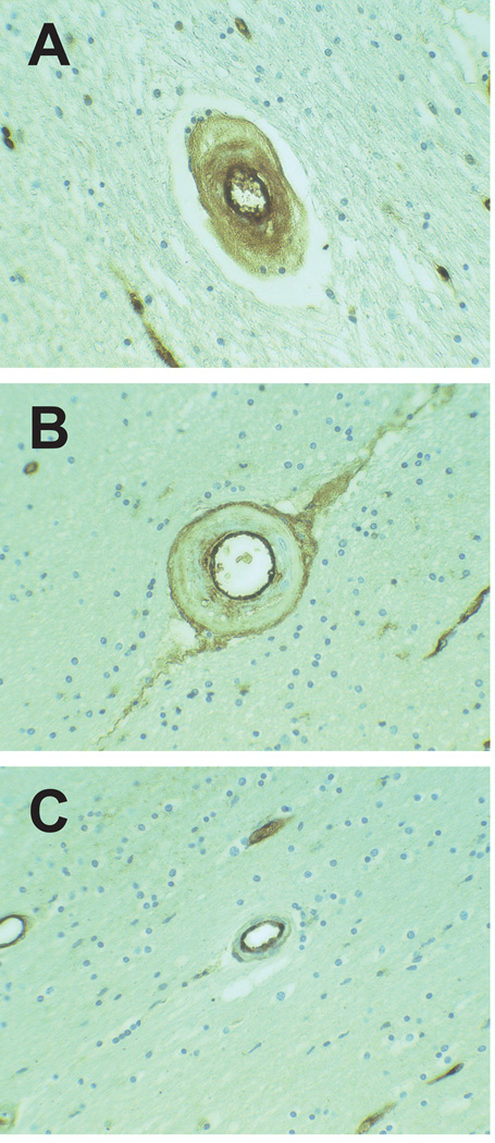
Figure 1.
Novel patterns of vascular vWF distribution in brain vasculature. Transmural distribution of vWF was very common in CADASIL small vessels (A). Both CADASIL and control samples contained double-barreled vWF profiles; the example shown is from a CADASIL brain (B). vWF was confined to the endothelium in vessels of both CADASIL and control vessels (C). All vessels were magnified at 400x.

