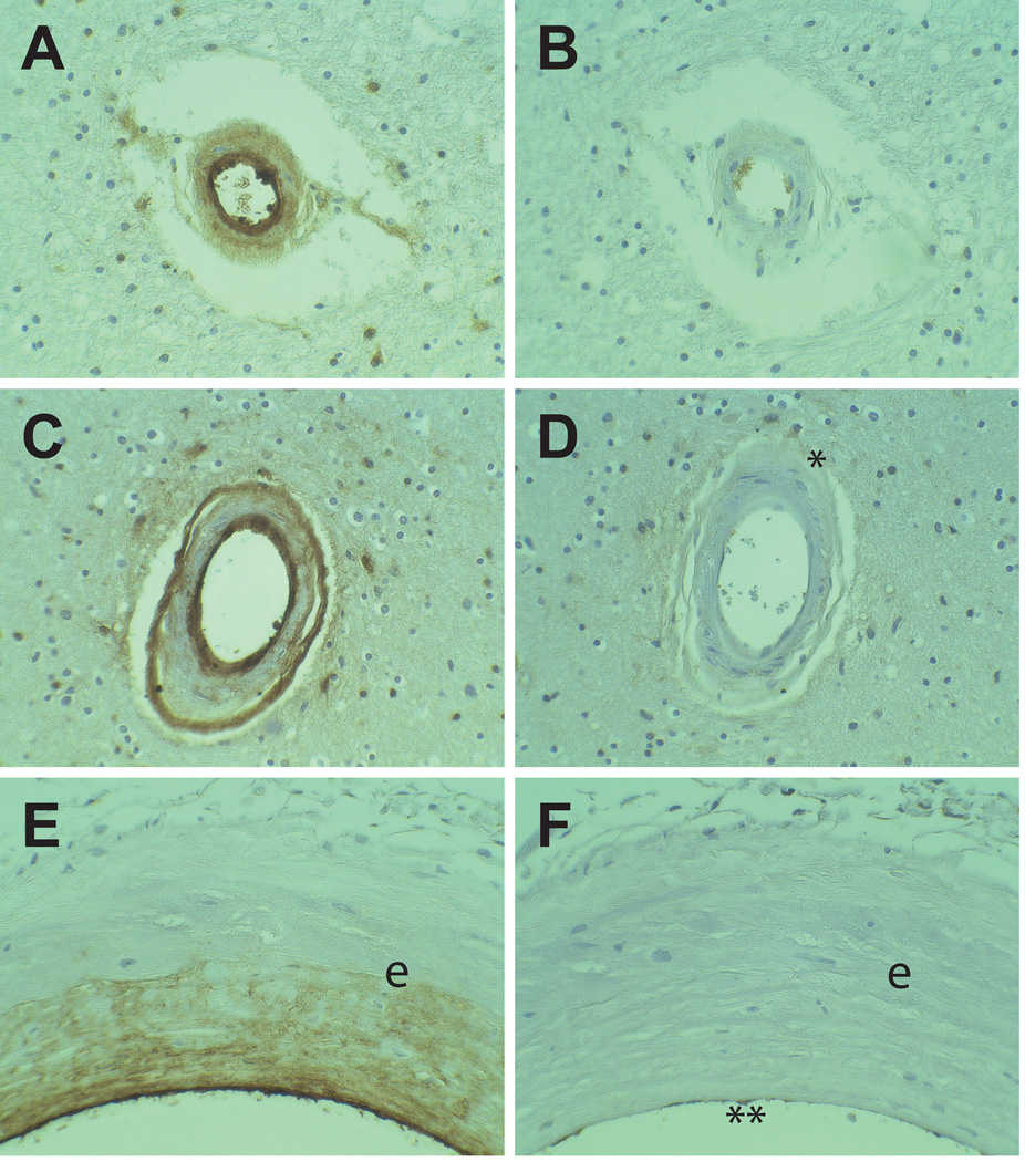
Figure 2.
Comparison between vWF and IgG immunoreactivity CADASIL brains. Serial sections from CADASIL brains were stained for vWF (A, C, E) and IgG (B, D, F). Small penetrating white matter arteries (A–B) with transmural deposition of vWF did not contain IgG. Arteries which exhibited a double barreled vWF staining pattern (C–D) faintly stained for IgG in the adventitia (*). Meningeal arteries (E–F) demonstrated heavy intimal deposition of vWF without IgG labeling. Endothlium of the arteries contained both vWF and IgG (marked ** in (F)). The elastic lamina is marked (e). All vessels were magnified at 400x.

