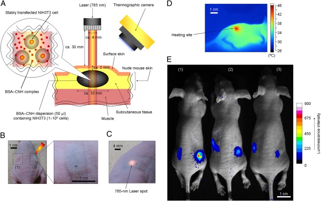
Fig. 3.
In vivo gene expression by laser-induced CNH complexes. (A) Location and geometry of suspensions consisting of NIH 3T3 cells and CNH complexes inside the tissue. (B) Photographs of suspensions consisting of NIH 3T3 cells and CNH complexes injected in a nude mouse. (C) Photograph of a 785-nm laser spot on the body of a mouse. (D) Thermographic measurement on the mouse’s body surface with 785-nm laser-induced CNH complexes [laser power, 150 mW (∼12 mW/mm2); irradiation time, 5 min]. (E) Bioimaging of in vivo gene expression driven by photothermal properties of CNH complexes. The right side of the body surfaces of mice were irradiated with a 785-nm laser at three preset temperatures: (1) 42 °C, (2) 39 °C, and (3) 45 °C.

