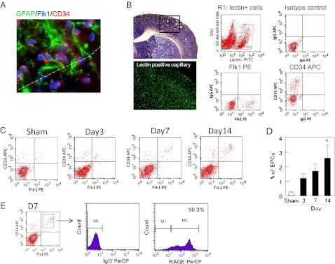
Fig. 3.
Accumulation of EPCs in the peri-infarct cortex during stroke recovery after transient focal cerebral ischemia in mice. (A) At 14 d poststroke, Flk1 and CD34 double-positive cells were found in close proximity with GFAP+ reactive astrocytes. (Magnification, 40×.) (B) Endothelial cells in the brain were labeled by lectin infusion, and Flk1/CD34 double-positive cells in the lectin-positive endothelial population were operationally quantified as EPCs with FACS. (Magnification, 10× for imaging lectin positive capillary.) (C) Representative FACS distributions of EPCs from 3 to 14 d after ischemic onset. (D) FACS quantitation showed that EPCs were steadily increased in the peri-infarct cortex over the 14-period of stroke recovery. *P < 0.05 vs. sham-operated group. (E) Further FACS analysis confirmed that these EPC subsets were also positive for the HMGB1 receptor RAGE.

