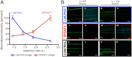
Fig. 3.
Staining of specific phage clones correlates with Fn fiber strain. Phage clones displaying the LNLPHG and RFSAFY peptides were labeled with AlexaFluor 633 SE and incubated on Fn fibers (1 × 10¹¹ phage per sample). (A) Staining of labeled LNLPHG phage decreases with increasing fiber strain, and staining of RFSAFY phage increases with increasing fiber strain. (B, i–xii) Fluorescence images of labeled phage clones on Fn fibers under varying strain. LNLPHG phage binding to relaxed (λ = 0.93) (i, ii) and strained (λ = 2.64) Fn fibers (iii, iv). RFSAFY phage binding to relaxed (λ = 0.93) (v, vi) and strained (λ = 2.64) Fn fibers (vii, viii). Labeled control phage show minimal binding (ix–xii). Error bars are SD. All images acquired with 63× oil immersion objective. (Scale bar: 20 μm.)

