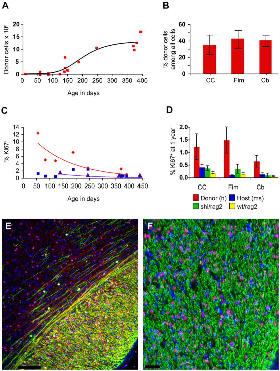
Figure 6. Long-term survival was associated with humanization of the recipient white matter.
A. Perinatally transplanted human GPCs increase in number asymptotically over the course of a year. From an initial dose of 300,000 on postnatal day 1, the cells increase to an average of 12 million/brain by one year post transplantation (y = −9,898,000 + 733,632×+ (−5709)2; r2 = 0.83).
B. By one year, donor cells comprised over 40% of all cells in the fimbria and cerebellar white matter, and over a third in the corpus callosum. Since the total cell count includes host vascular cells and microglia, the human donor-derived cells appeared to comprise a net majority of all glial cells by that stage. C. Over the year after implantation, the rate of human GPC proliferation in white matter declined exponentially (red; y = 14.013e−0.0475×; r2 = 0.79). At 8 weeks, 12.35% of human GPCs in the mouse corpus callosum are Ki67 positive, but by one year, an average of 1.22% are Ki67%. From 5 to 12 months, the percentage of Ki67+ mouse cells in the corpus callosum of untreated rag2 null mice also declines exponentially, but beginning at a lower rate (purple; y = 2.9154e−0.0497×; r2 = 0.83). The Ki67+ percentages of endogenous mouse cells in the same sections of transplanted mice from which the hGPC percentages were obtained, however, do not follow a pattern of exponential decline (blue; y = 1.3684e-0.02× r2 = 0.1855). D. At one year, the percentage of hGPCs in white matter that are Ki67+ (red) exceeds that of the endogenous mouse cells in the same mice (blue), as well as that of untreated rag2 null mice (yellow), and that of 4 month old untreated shiverer/rag2 homozygotes (green) in corpus callosum, fimbria and cerebellum.
E–F. Progressive myelination (MBP, green) of mouse axons (neurofilament, in red) was attended by chimerization of the recipient white matter, such that by 20 weeks, host cells (DAPI, blue) are exceeded by human donor cells (human nuclear antigen, hNA, purple, as blue co-labeled with hNA, red). E. Parasagittal section including dorsal callosum and overlying cortex of a transplanted shiverer at 20 weeks, showing human donor-derived myelination of callosum, and admixture of host (blue, DAPI) and donor cells (purple, as blue co-labeled with hNA, red). Both myelinated (MBP, green) and unmyelinated (NF, far red) fibers are evident traversing lower cortical layers. F. Higher magnification section through fimbria of hippocampus, showing myelinated fibers viewed en face, with admixed mouse (blue) and human (purple, representing blue co-labeled with hNA, red) cells. Scale: E, 100 μm; F, 50 μm.

