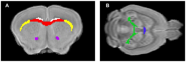
Figure 1.

Regions of interest (ROI) for diffusion measurements. Non-diffusion weighted images of a mouse brain in (A). Coronoal view of brain with ROI for corpus callosum (red), external capsule (yellow), cingulum (white), and anterior commissure (purple) or (B). Horizontal view with ROI for dorsal hippocampal commissure (green) and ventral hippocampal commissure (blue).
