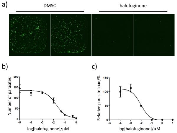
Figure 2.
Halofuginone inhibition of P. berghei parasite load in liver cells. Visualization of P. berghei sporozoite load in HepG2 cells by staining with an antimalarial antibody (A). Cells were infected in the presence of DMSO or 1 μM halofuginone and fixed 36 – 48 hours after sporozoite addition. Dose-response curves of parasite load assessed by quantification of parasite numbers with antibody staining (B) and by relative luminescence signal after infection with a transgenic luciferase reporter strain of P. berghei sporozoites (C) yield IC50 values of 17 ± 8 nM and 7.8 ± 3 nM, respectively. Data are shown as the mean ± the standard deviation.

