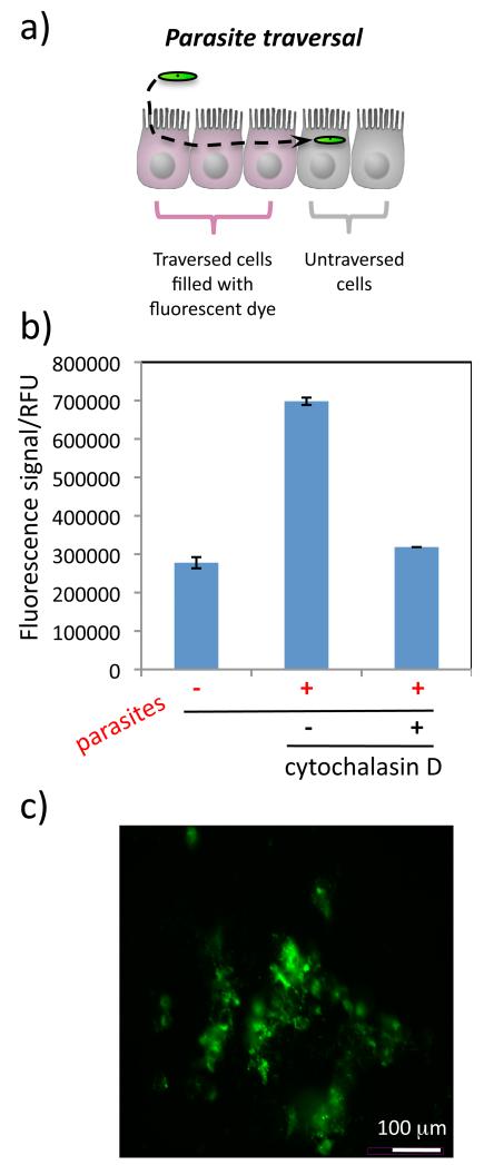
Figure 4.
Model of sporozoite traversal through cells (A). Within 2 hrs HepG2 cells that have been traversed by P. berghei sporozoites (in green) fill with Rhodamine Green dextran that was added to the cell medium (cells in pink). The relative numbers of traversed HepG2 cells can be measured using a fluorescence plate reader (B). Data are shown as the average fluorescence reading of 4 measurements on the same 384-well plate and error bars show the standard deviation. Sporozoites were incubated with cytochalasin D before addition to the cells as a control. Visualization of samples with a fluorescence microscope confirms that the fluorescently labeled dextran has entered a population of HepG2 cells (C).

