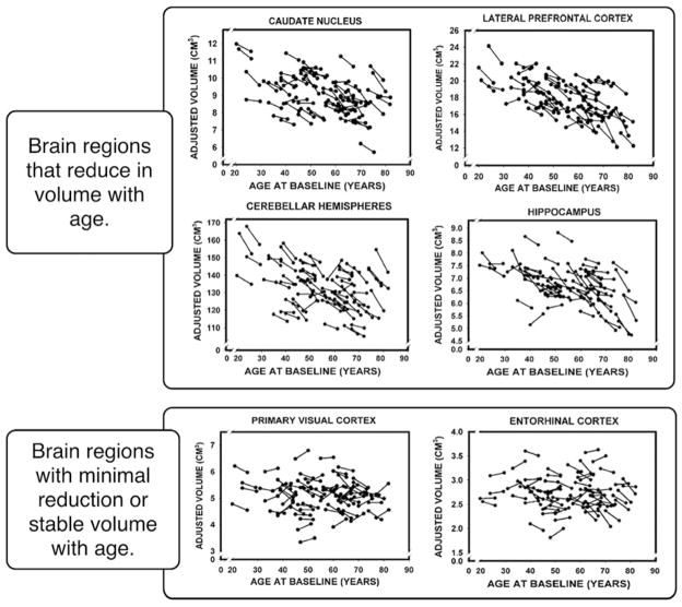
Figure 2.
Cross-sectional and longitudinal aging brain volumes across various brain regions (adapted from Raz et al. 2005). Each pair of line-connected dots represents an individual subject’s first and second measurement. The caudate, hippocampal, cerebellar, and frontal regions all show both cross-sectional and longitudinal reduction in volume with age. The entorhinal, parietal, temporal, and occipital regions are relatively preserved with age.

