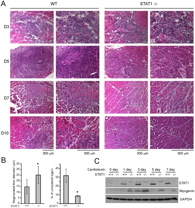
Figure 1. Muscle regeneration was accelerated in STAT1−/− mice after cardiotoxin injection.
Tibialis anterior muscles from 2-month-old wild-type and STAT1−/− mice were injected with 30 ul of 10 nM cardiotoxin. The TA muscles were harvested at different time points as indicated after injury. (A) The muscles were fixed in 4% paraformaldehyde/PBS (pH 7.4) for 1 hour at 4°C, infiltrated sequentially with 10%, 20% and 30% sucrose, and embedded in O. C. T. solution. Six micrometers of cryosections were stained with hematoxylin and eosin. Bars: 300 um. (B) The diameter of the regenerated fibers and the percentage of un-repaired region from three independent experiments were measured 10 days post-injury. Data were presented as mean ± SD. Asterisk: p<0.05. (C) Tibialis anterior muscles were homogenized in protein lysis buffer. Equal amounts of protein were separated by SDS-PAGE followed by western blotting with different antibodies as indicated.

