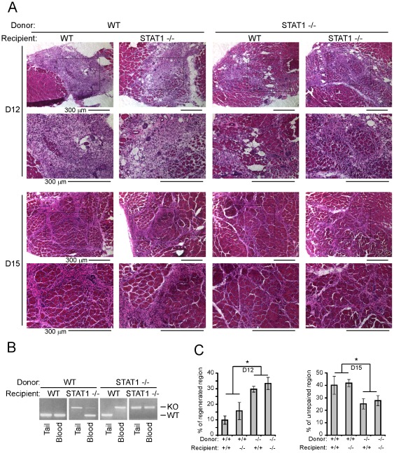
Figure 2. Loss of STAT1 in the bone marrow-derived cells accelerated muscle regeneration.
Donor bone marrow cells were freshly prepared from the femurs of donor mice and filtered through a sterile 40 µm nylon Cell Strainer to remove debris. The cells were suspended in DMEM without serum prior transplantation. The recipient mice were given 8 Gy Gamma-irradiation and each injected with 2×10⁶ donor bone marrow cells through retro-orbital vein. Six to eight weeks after bone marrow transplantation, tibialis anterior muscles of the recipient mice were injected with 30 ul of 10 nM cardiotoxin. (A) The muscles were fixed in 4% paraformaldehyde/PBS (pH 7.4) for 1 hour at 4°C, infiltrated sequentially with 10%, 20% and 30% sucrose, and embedded in O. C. T. solution. Six micrometers of cryosections were stained with hematoxylin and eosin. Bars: 300 um. (B) The tails and peripheral blood cells from the recipient mice were harvested, and genomic DNA was extracted. PCR was used for genotyping. The upper and lower bands indicate genotypes of STAT1−/− and wild-type respectively. (C) The percentage of regenerated region 12 days post-injury and unrepaired region 15 days post-injury from three independent experiments were measured. Data were presented as mean ± SD. Asterisk: p<0.05.

