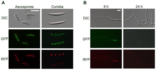
Figure 7. Cellular localization of MYT2.
MYT2 was fused with green fluorescent protein (GFP), and histone H1 was fused with red fluorescent protein (RFP). Co-localization of MYT2-GFP and hH1-RFP in spores (A) and germinated conidia 9 and 24 h after inoculation in complete medium (B). DIC, differential interference contrast Scale bar = 20 µm.

