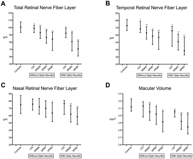
Figure 1. Retinal Axonal Degeneration in Multiple Sclerosis is Increasingly Prominent in More Advanced Stages of Disease and Proportionally Greater in Eyes Previously Affected by Symptomatic Optic Neuritis.
Retinal Nerve Fiber Layer thickness (A, B, C) and macular volume (D), as measured by spectral-domain optical coherence tomography (Heidelberg Spectralis) in a cross sectional sample of patients with high-risk Clinically Isolated Syndromes (CIS) (n = 45), Relapsing-Remitting MS (RRMS) (n = 403), Secondary-Progressive MS (SPMS) (n = 60), Primary-Progressive MS (PPMS) (n = 33) and unaffected controls (n = 54). Both the total and temporal peripapillary RNFL were thinner in CIS patients compared to controls in eyes without prior symptomatic optic neuritis. RNFL measures were nearly identical between SPMS and PPMS patients in eyes without optic neuritis, but macular volumes were lower in PPMS compared to SPMS patients in eyes without optic neuritis (p<0.05). The black dots denote the median, and the bars signify the interquartile range. *p<0.05, **p<0.001 refers to the comparison with unaffected controls using linear regression to adjust for age and sex.

