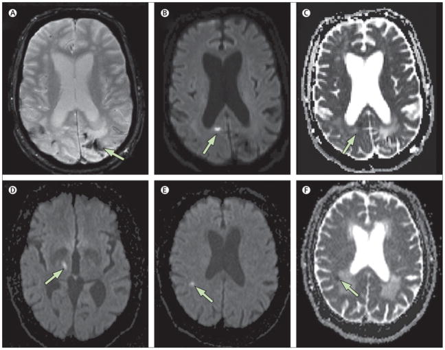
Figure 3. MRI Diffusion-Weighted Imaging of Small Acute Infarction (Sample).
Example of small acute areas of restricted diffusion detected incidentally on MRI. Top panels (A–C): A 70 year man with cerebral amyloid angiopathy who underwent MRI as part of a research study. Hemosiderin staining from prior hemorrhages is seen on the T2*-weighted gradient-recalled echo (GRE) sequence (A). Separate from these prior hemorrhages, an asymptomatic small hyperintensity is seen in the right occipital cortex on the diffusion-weighted image (DWI, panel B) with evidence of restricted diffusion on the apparent diffusion coefficient sequence (ADC, panel C), consist with an acute small cortical infarct. Bottom panels (D–F): A 67-year old man with an acute symptomatic lacunar infarct in the right thalamus (D, DWI sequence) also demonstrates an asymptomatic small 4.5 mm lesion in the right parietal white matter that is hyperintense on DWI (panel E) with restricted diffusion on ADC (panel F). An asymptomatic simultaneous small vessel infarct was suspected because no proximal source of embolism was identified and there was evidence of coexisting chronic cerebral small vessel disease (note confluent white matter lesions exhibiting increased diffusion in panel F).

