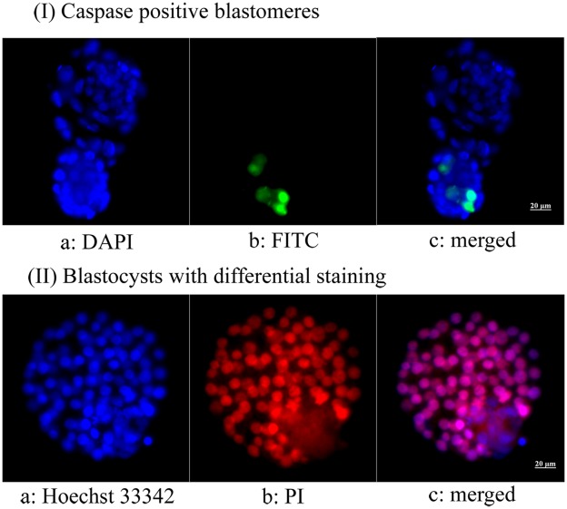
Figure 1. Microphotographs showing fluorescent caspase staining and differential staining of blastocysts derived from vitrified-warmed immature mouse oocytes.
(I) Apoptotic blastomeres appear green; (II) red-pink indicates trophectoderm and blue indicates inner cell mass (400×). (I) a: DAPI, b: FITC, C: merged; (II) a: Hoechst 33342, b: propidium iodide, c: merged.

